
Supplemental movies to my PhD thesis Sensing Cilia
Chapter 3 - Imaging adult C. elegans live using light sheet microscopy
Movie S3.1: IFT dynamics.
IFT dynamics of OSM-6::EGFP, an IFT-B particle complex component, in the phasmid cilia of C. elegans, imaged with light-sheet microscopy. Related to Figure 2D.

Movie S3.2: Head region neurons with light-sheet microscopy.
Movie of z-stack of neuronal microtubules, visualized with TBB-4::EGFP, imaged with light-sheet microscopy. Related to Figure 3A.

Movie S3.3: Head region neurons with epifluorescence microscopy.
Movie of z-stack of neuronal microtubules, visualized with TBB-4::EGFP, imaged with epifluorescence microscopy. Related to Figure 3C
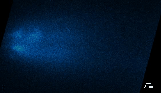
Movie S3.4: Neuronal cell bodies in the head region.
3D visualization of z-stack of GCaMP, imaged with light-sheet microscopy. Related to Figure 4A.

Movie S3.5: Neuronal activity in the tail region.
Neuronal activity visualized with GCaMP in the tail region, imaged with light-sheet microscopy. Related to Figure 4B-E.

Chapter 4 - Cutting off ciliary protein import:
Intraflagellar transport after dendritic femtosecond-laser ablation
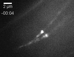
Movie S4.1: IFT-dynein redistribution and slow-down.
Fluorescence movie of pair of phasmid cilia (XBX-1::EGFP strain) pre and post-ablation. Time and scale bar are indicated. Same data as in Figure 4.2.
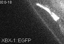
Movie S4.2: IFT dynein and OSM-3 distribution and dynamics in C. elegans phasmid cilia before and after low-concentration azide treatment. Sodium azide is added at t = 0 s. Scale bar, 2 um. Video plays at 5x real time (time indicated). Video corresponds to Figure4.4A, third horizontal panel, and Supplemental Figure 4.2A, third horizontal panel.

Chapter 5 - Single-molecule turnarounds of intraflagellar transport
at the C. elegans ciliary tip

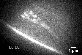
Movie S5.1: Single-Molecule Tip Turnarounds of OSM-6::EGFP in C. elegans Phasmid Cilia. Related to Figures 5.1 and 5.2. Ciliary bases are located in the lower half of the video, ciliary tips in the upper half.

Movie S5.2: Transient Accumulation of OSM-6::EGFP at the Ciliary Tip. Related to Figure 5.1. Ciliary base is located in the lower half of the video, ciliary tip in the upper half.
Chapter 6 - OCR-2 is distributed along C. elegans chemosensory
cilia by diffusion in a local interplay with intraflagellar transport

Movie S6.1: Redistribution of IFT-dynein upon repellent exposure. Fluorescence microscopy image sequence showing the reversible redistribution of IFT-dynein (XBX-1::EGFP) in the distal segments of the phasmid chemosensory cilia of C. elegans after the addition of a droplet of 5 µl 0.1% (W/V) SDS. Related to Figure 6.1.
Movie S6.2: Single-molecule imaging of SRB-6.
Single-particle fluorescence image sequences of endogenously labeled SRB-6, showing its low expression level in the phasmid cilia and mostly diffusive and salutatory motility.

Movie S6.3:Single-molecule imaging of OCR-2.
Collage of example single-molecule image sequences of OCR-2::EGFP, demonstrating the diversity in OCR-2 motility. Related to Figure 6.3A.